

Equipment
Hardware
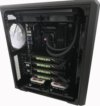
LYAX Workstation
 The image processing workstation is configured with 2 Intel Xenon i5 processors with 8 cores (2.9 Ghz) and 256 GB of RAM (DDR3 memory). The workstation is ideal to run computational processed that require high amount of memory such as ilastik pixel classification or autocontex.
Room 001 (Booking calendar)
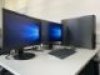
MiFCOM Workstation 2018
The image processing workstation is configured with new generation of fast processors like Intel Core i9 with 10 cores (3.0-3.5 GHz) and an optimal amount of RAM (128 GB DDR4 memory). To gain the best performance from Arivis Vision4D and InViewR (VR), the workstation is equipped with 1 x NVidia GeForce TitanX Pascal graphic cards (12 GB VRAM per card).
Room 001 (Booking calendar)
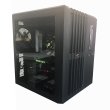
MiFCOM Workstation 2017
The image processing workstation is configured with new generation of fast processors like Intel Core i7 with 10 cores (3.3 GHz) and an optimal amount of RAM (128 GB DDR4 memory). To gain the best performance from Arivis Vision4D and the deep learning machine, the workstation is equipped with 4 x NVidia GeForce TitanX Pascal graphic cards (12 GB VRAM per card).
Room 001
Dell PowerEdge T630 GPU Server
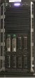
The server is configured with 2 x 10 core Intel Xeon processors and 1TB of RAM. The server runs Huygens SVI deconvolution software (hosted by Nikon Imaging Center), ImageJ/Fiji and ilastik. Heidelberg University life scientists can have access to the server and all the software installed from any workstation located at the campus upon previous registration here.
Oculus Rift
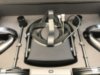 Virtual reality (VR) systems are becoming more and more powerful, because they allow rendering and processing of large 3D and 4D (3D over time) biomedical image data from different perspectives. The oculus rift system can be use to navigate interactively through microscopic data in VR.
Software
Open Source Software
The Math-Clinic core facility offers support in bioimage data processing and analysis using different open source software. Here you can find an useful list of the most common open source software for bioimage data analysis:
Dr Carlo Beretta offers his help to those scientists interested in learning how to model cytoskeleton structures using the simulation software CYTOSIM (Nedelec F, New Journal of Physics 9 (11) 427, Nov 2007).
Commercial Software Solutions at the Core Facility
SVI Huygens Software for Image Deconvolution
Deconvolution of image data stack improves contrast and resolution and is especially useful for colocalization analysis and 3D rendering. The Math-Clinic in collaboration with the Nikon Imaging Center at Heidelberg University offers Huygens software for deconvolution. Heidelberg University members can access to Huygens deconvolution software after previous registration at Huygens Remote Manager (HRM). The software is hosted on the Math-Clinic / Nikon Imaging Center server and registered users can get access to the program remotely using the HRM web interface. On request, users can get a short introduction to the program. Access to deconvolution software is free of charge for all CellNetworks members. Others may get access to Huygens for a small fee of 80 € per year and group.
NIS-Elements is the central solution for image acquisition and analysis offered by Nikon. It handles multi-dimensional imaging flawlessly, with support for capture, display, peripheral device control, data management and analysis of up to six dimensions (X,Y,Z, Lambda (wavelength), T, multipoint). It also offers sophisticated image processing features, such as an extremely powerful deconvolution module, an intuitive tool to design bioimage analysis workflows and, an exclusive one-click database capability. The software can be use free of charge.
The Arivis Vision4D software is the state-of-the-art software to handle multidimensional large data sets. The software supports most microscopy image formats and performs interactive rendering without RAM constrains. The software has been purchased by CellNetworks Math-Clinic core facility and the Nikon Imaging Center (jointed application 2017 CellNetworks Equipment Program). Life science groups of Heidelberg University and Medical Faculty can use the software free of cost; a small fee can be applied to external institutions (DKFZ, MPI and EMBL).
Arivis InViewR is a newly in rapid development virtual reality (VR) software used to visualize and process large biological images. The software uses the VR Oculus Rift system (already purchased by the Math-Clinic) to navigate interactively through microscopic data and manually or semi-automatically segment regions of interest. Arivis InViewR can be used to render many microscopy images such as LSM, confocal (CLSM), 2-PM, electron microscopy, computer tomography and magnetic resonance tomography data. The software has been purchased by CellNetworks Math-Clinic core facility and Prof Dr Thomas Kuner (jointed application 2018 CellNetworks Equipment Program). Life science groups of Heidelberg University and Medical Faculty can use the software free of cost; a small fee can be applied to external institutions (DKFZ, MPI and EMBL).
Please contact Dr Carlo Beretta if you are interested in using the software.